The heart is a muscular organ that collects blood that is deoxygenated blood from every area of the body and transports it to the lungs where it is oxygenated and carbon dioxide is expelled.
The blood is then transported from the lungs and distributed to every region of the body.
Around 7,200 liters of blood are circulated by the heart each day throughout the body.
The center of the chest is where the heart is located, and it leans slightly to the left.
The heart typically beats 100,000 times per day or nearly 3 billion times throughout a lifetime.
The heart of an adult beats between 60 and 80 times per minute, but the heart of a newborn baby beats between 70 and 190 times per minute, which is quicker than the heart of an adult.
In this article, we’ll discuss the various components that make up the human heart while also providing a quick overview of each component’s characteristics and functions.
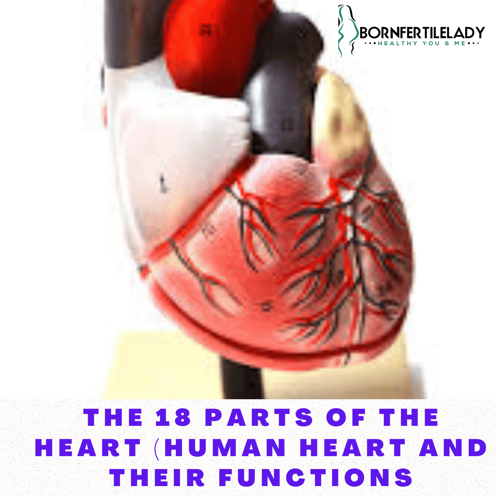
The 18 parts of the human heart and their functions
Myocardium
The layer of contractile muscle known as the myocardium is responsible for the mechanical process of pumping blood into the body.
It is a muscle that works automatically, or without the assistance of a neurological impulse from the nervous system. The heart alone produces the contractile signal.
The ventricles, which make up the lower portion of the heart, have more myocardium than the atria, which make up the upper portion.
Additionally, it is more common on the left than the right side of the heart.
Endocardium
The lining of the various heart chambers is called the endocardium.
Similar to the other blood vessels, it is primarily made up of endothelial cells and has a thin layer of lax connective tissue.
Pericardium
The pericardium is a fibrous membrane that isolates and protects the heart as well as the large blood arteries that attach to it.
The pericardium, which has two layers—serous pericardium and fibrous pericardium—can be compared to a large bag.
Because of its viscosity and capacity for protection, the heart can beat freely without being hampered by neighboring structures
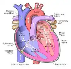
Right Auricle
The cavity in the upper-right region of the heart, above the right ventricle, is known as the right atrium.
Both the superior and inferior vena cava connect to it bringing the blood that has already been circulating through the body—which is low in oxygen and high in carbon dioxide—to the heart. Before entering the right ventricle, the blood enters the right atrium.
Right ventricle
The right atrium, from which the right ventricle receives blood, is connected to it.
The pulmonary artery, which connects the right ventricle to the lungs where gas exchange takes place, is in charge of transporting blood from the right ventricle to the lungs.
Before the left ventricle, it slightly contracts.
Tricuspid valve
The valves are located between the atria and the ventricles as well as between the ventricles and the arteries.
Their primary purpose is to prevent blood reflux and to segregate the atria and ventricles; they are composed of connective tissue.
They only become perfused with blood, and the myocardium’s contractions cause them to immediately close after opening.
The valve between the right ventricle and the right atrium is known as the tricuspid valve.
Because it is made up of three sheets of connective tissue and is connected to the right ventricle by tendons and tiny papillary muscles, it is known as the tricuspid valve.
Pulmonary valve
The valve that joins the right ventricle and pulmonary artery is known as the pulmonary valve.
The right ventricle contraction causes it to open, allowing blood to enter the artery.
Left Auricle
The pulmonary veins supply blood to the left atrium (there are 4, 2 coming from the right lung and 2 coming from the left lung).
Through the mitral valve, blood enters the left ventricle from the left atrium.
Left ventricle
The strongest heart muscles are found in the left ventricle.
Blood is pumped out of this ventricle and into the aortic artery, which divides to supply the remaining blood in the body.
This ventricle must produce blood pressure that is substantially higher than that of the right ventricle.
Mitral valve
The left atrium and left ventricle are divided by the mitral valve.
It is connected to the ventricle with tendons and papillary muscles, just like the tricuspid valve, which prevents it from opening both when the ventricle is empty and when the ventricle contracts.
Aortic valve
The valve that separates the aortic artery from the left ventricle is known as the aortic valve.
It must be resilient to high blood pressure.
Tendon cords
Tendon cords, which are made of connective tissue, link papillary muscles to the mitral and tricuspid valves.
Papillary muscles
The wall of the ventricles contains the papillary muscles.
Their primary purpose is to keep the valves closed and stop blood from refluxing or flowing backward into the heart circuit. They have the shape of a cone.
Sinoatrial node
A sinoatrial node is a group of cells that serves as our heart’s natural pacemaker by signaling when it needs to contract.
Muscle contraction is brought about by an electrical signal that originates in the sinoatrial node and passes via specific structures as it descends the heart.
Atrioventricular node
Close to the sinoatrial node is the atrioventricular node.
It can also act as the heart’s pacemaker, however, it only does this in cases where the sinoatrial node fails.
Less often than the sinoatrial node, the atrioventricular node produces impulses.
Atrioventricular fascicule
The excitatory impulse from the nodes passes from the atria to the ventricles by a muscular tissue called the atrioventricular fascicule, also referred to as the bundle of His.
Coronary arteries
The coronary arteries encircle the heart and supply the organ with nourishment.
These divide into the left and right coronary arteries, each of which primarily supplies blood to the respective half of the heart.
The most prevalent kind of heart disease is a disease of the coronary arteries.
Read also: 10 Health Benefits of Having Sex During Pregnancy (is it safe?)
Coronary veins
The coronary veins are responsible for bringing blood low in oxygen from the heart and its surrounding tissues back to the heart.
Surprisingly, these veins efficiently and elegantly drain right into the atrium.